
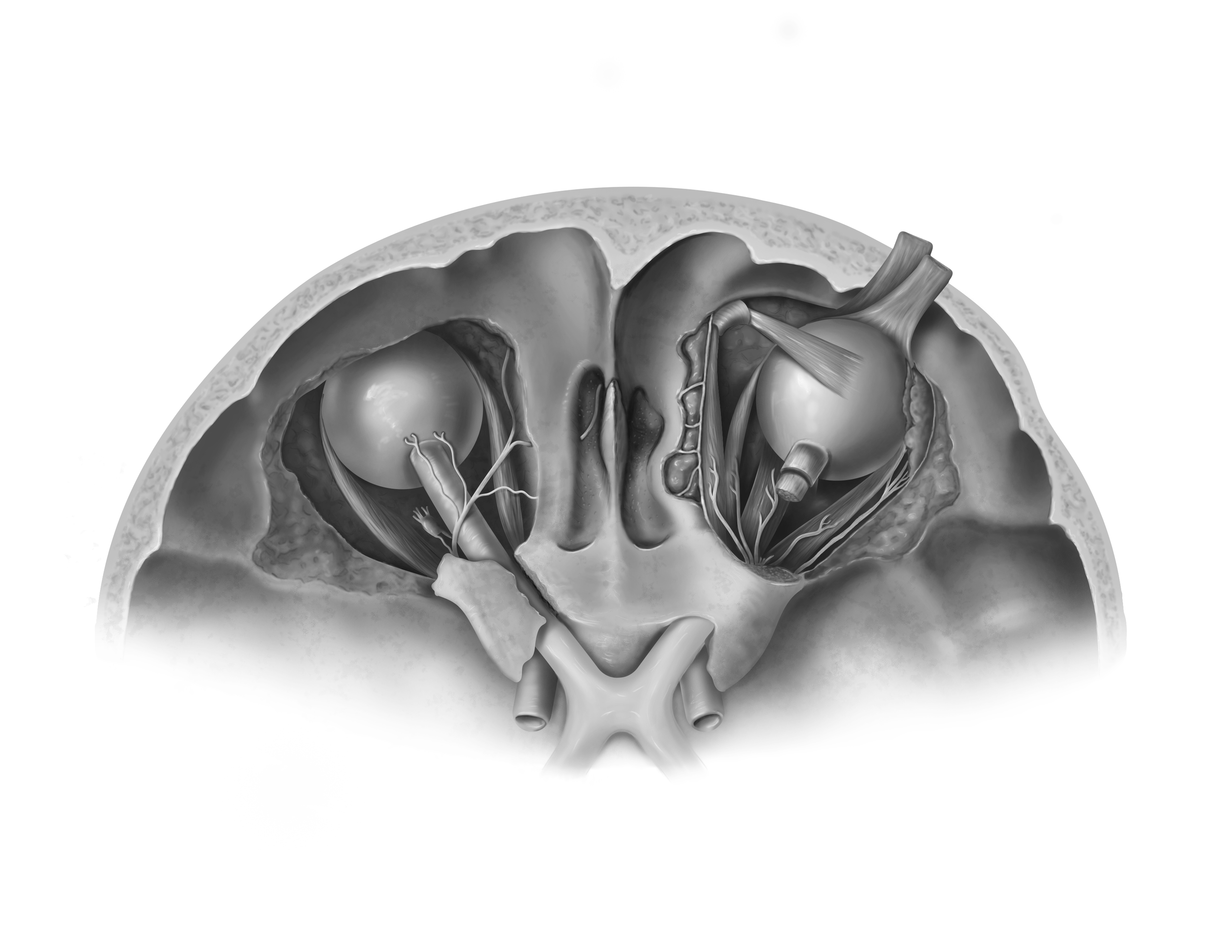
Anatomical Illustration: Superior Orbital Cavity
Audience: Anatomy Students
Client: Professor Dave Mazerski
Format: Labeled Anatomical Illustration
Tools used: Adobe Photoshop
Completed: December 2025
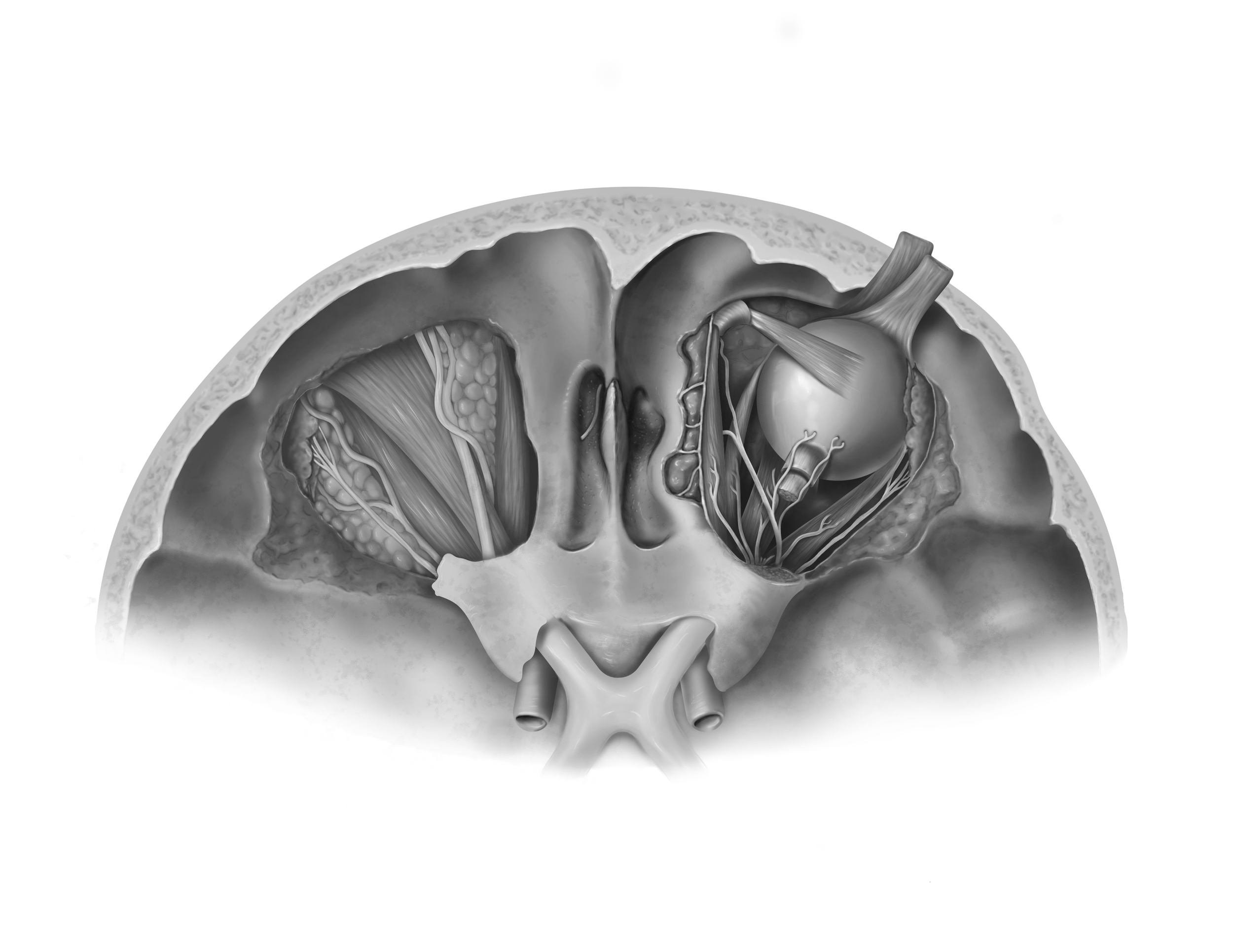
As a completion of my Human Anatomy studies within the MSc Biomedical Communications program, I created this raster illustration based on an orbital cavity dissection in Grant’s Museum at the University of Toronto. Two versions of this illustration were produced to maximize the amount of anatomical information that could be portrayed to the viewer. The first version of this illustration is a deep dissection into the eye sockets where lateral, medial, inferior muscles, and nerves of the eye can be seen. A second version of this illustration shows the superior muscles and nerves of the left eye.
Please check back later!
1. Sketching from Dissection
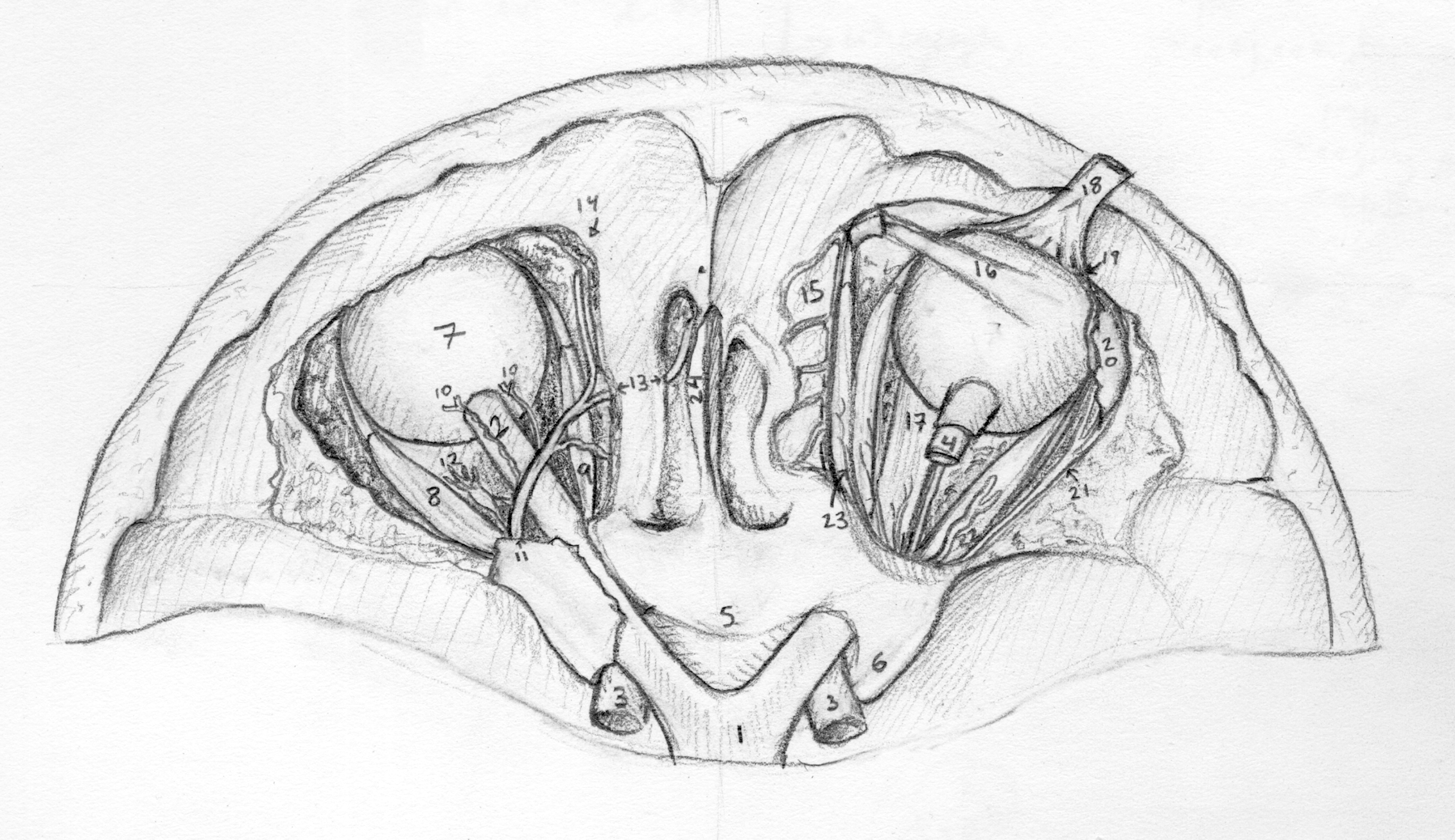
I created an initial sketch observing a prosection in Grant’s library at the University of Toronto. The prosection was dissected many years ago, so tissue was highly degraded. I used references from Grant’s Atlas while drawing the prosection to correct the tissue degradation.
2. Production Process
I created vector shapes of each major anatomical structure on Adobe Illustrator. By doing this, I could add shading to each object on Adobe Photoshop without worrying about drawing over the edge.
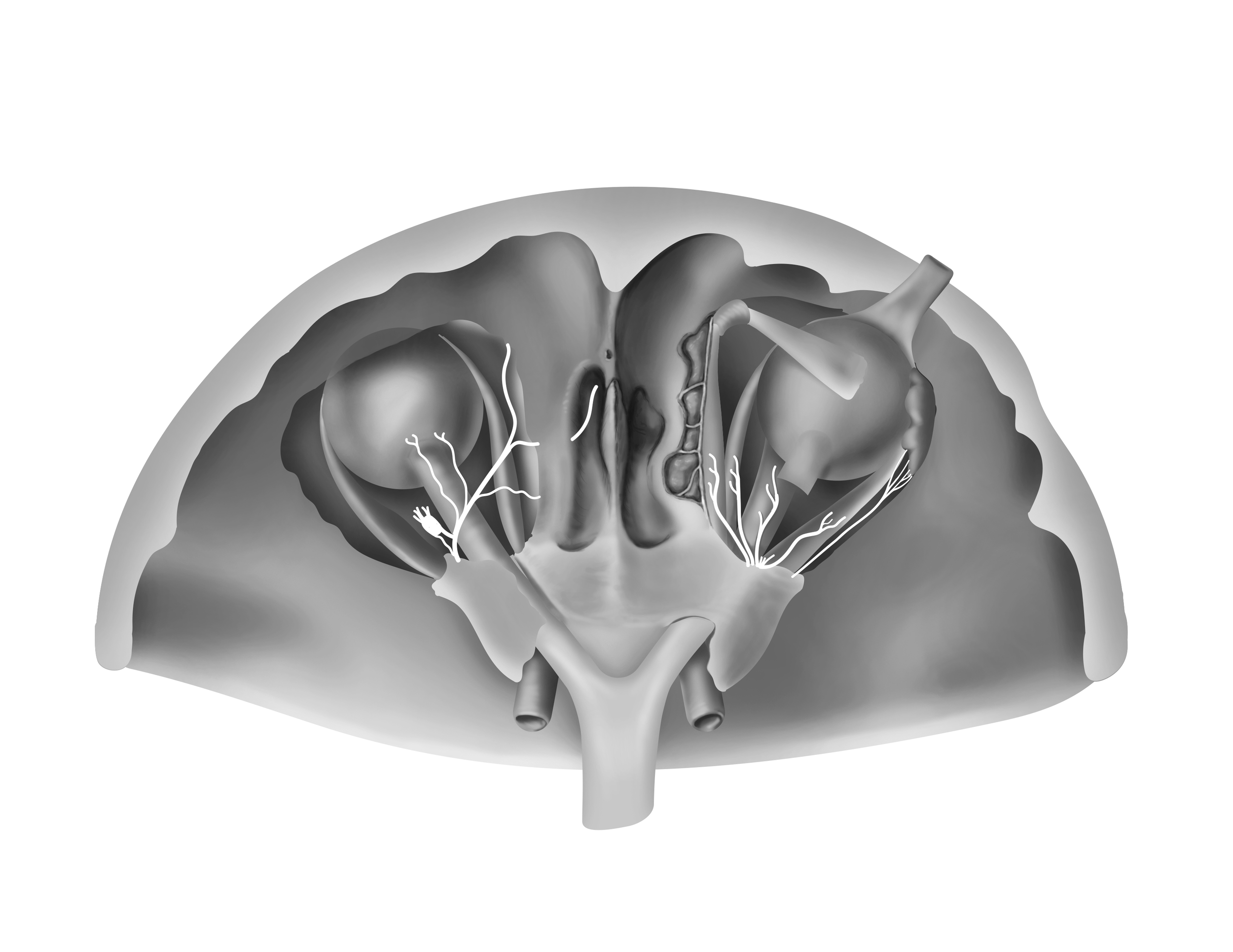
I also learned about the optic chiasm and corrected my anatomy as I went.
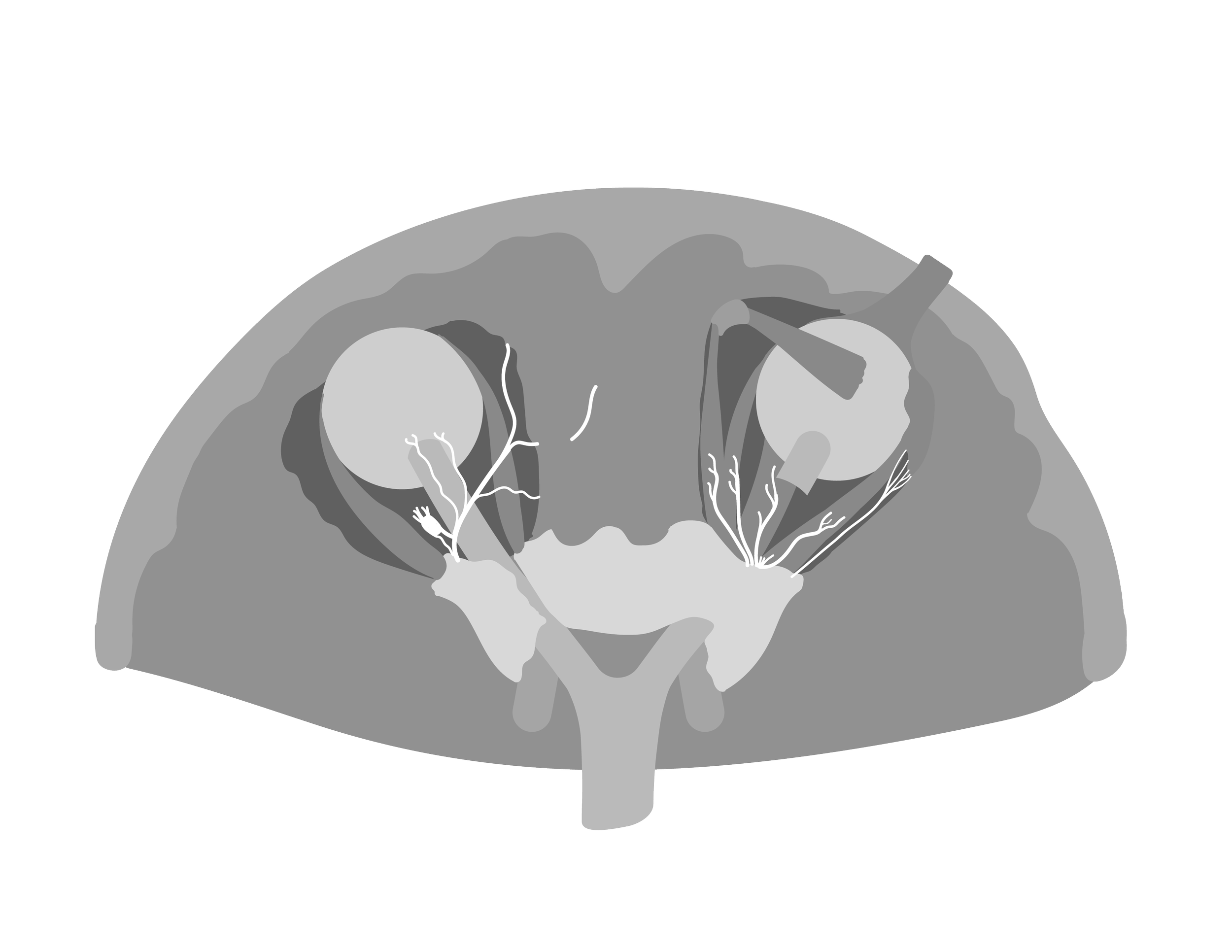
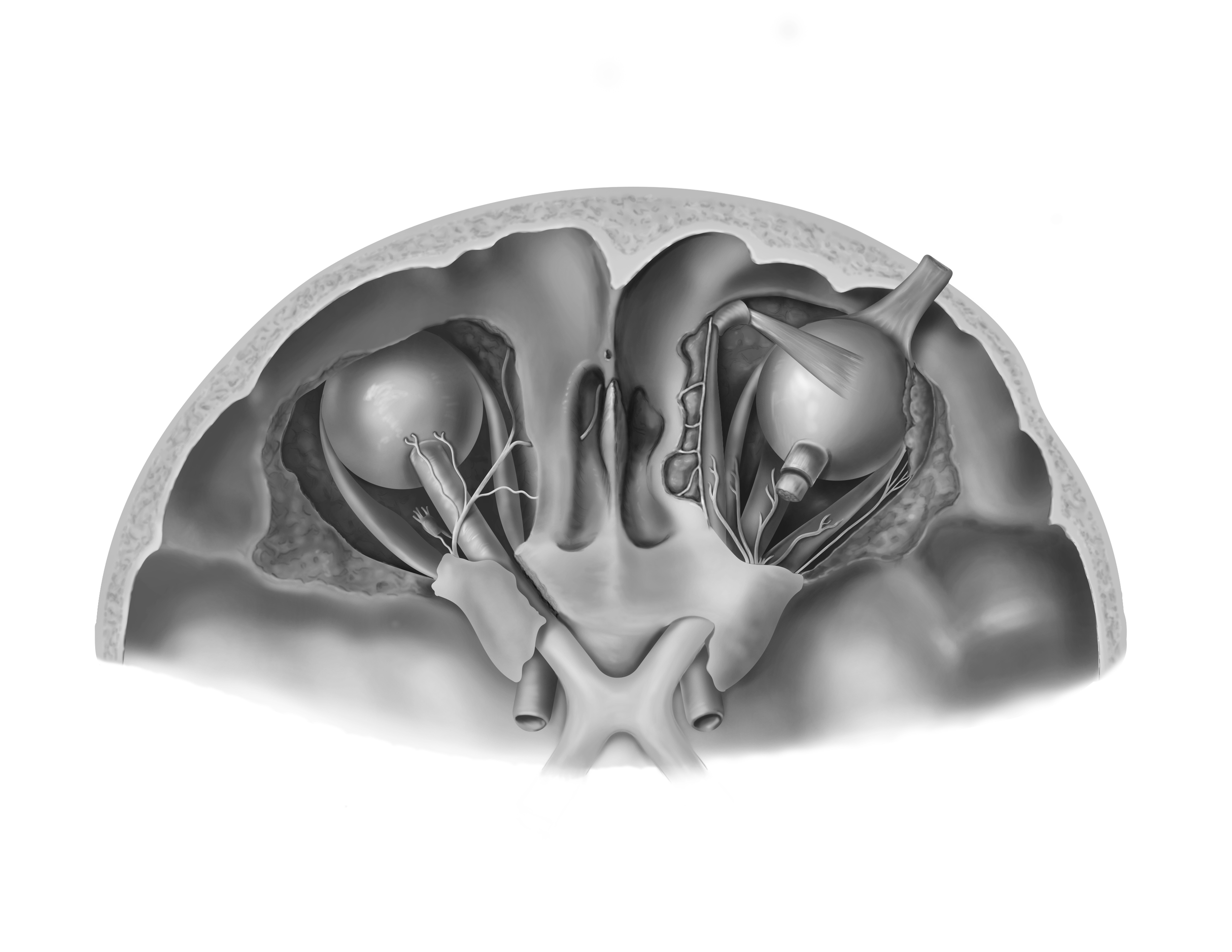
After some thought, I considered how many of the superficial muscles, nerves, and fat of the orbit were being excluded from my illustration. To maximize the anatomy I could depict in this illustration, I decided to illustrate one eye with a superficial dissection and one eye with a deep dissection.
References
Agur, Anne M. R., and Arthur F. Dalley II. Grant's Atlas of Anatomy. 14th ed., Wolters Kluwer, 2017.
Clemente, Carmine D. Anatomy: A Regional Atlas of the Human Body. 3rd ed., Urban & Schwarzenberg, 1987.
Schuenke, Michael, et al. Thieme Atlas of Anatomy. 3rd ed., Thieme Medical Publishers, 2021.